Table of Contents
Clinical Summary:
The Gap: Clinicians still triage lumbar spinal stenosis patients by MRI severity. The 2024 cohort data says that severity tells you less about who will respond to rehabilitation than almost anything else you can measure.
The Evidence: Shahidi and colleagues found that mild, moderate, and severe stenosis grades all show similar improvement patterns with supervised exercise. Nordsten-DS 5-year data (BMJ, 2024) confirms decompression alone matches decompression plus fusion. Surgical and conservative outcomes converge at 6–8 years.
The Takeaway: An adequate rehabilitation trial is the appropriate first step for almost every symptomatic stenosis patient, regardless of imaging grade.
A patient walks in with an MRI report that reads "severe central canal stenosis at L4-L5 with marked ligamentum flavum hypertrophy." She also walks the four blocks from the parking garage to your clinic without stopping. Her neighbor, with mild stenosis on imaging, can't make it to her own mailbox.
If you've been in clinic long enough, you've stopped being surprised by this. The MRI report and the patient in front of you don't always match. What's changed in the last three years is that the evidence finally caught up to what experienced clinicians have been seeing all along: imaging severity is a poor predictor of who responds to lumbar spinal stenosis rehabilitation.
Which means the algorithm a lot of us were taught (mild stenosis gets rehab, severe stenosis gets a surgical consult) is no longer defensible.
The Research Tension
The cleanest piece of evidence on this question comes from Shahidi and colleagues, who specifically asked whether radiographic stenosis severity predicts responsiveness to exercise-based rehabilitation. It does not. Mild, moderate, and severe grades all showed similar patterns of improvement when patients received appropriate intervention.
This is not an outlier finding. It sits alongside three other pieces of recent evidence that, taken together, restructure how stenosis decisions should be made:
- The Nordsten-DS five-year results (BMJ, 2024) found no difference between decompression alone and decompression plus fusion for degenerative spondylolisthesis. Oswestry Disability Index, leg pain, back pain, quality of life: all equivalent at five years.
- The convergence finding: surgical and conservative management produce statistically indistinguishable outcomes by 6–8 years across multiple trials. Surgery's short-term advantage is real, but it erodes.
- Wesselink et al. identified paraspinal multifidus intramuscular fat at 50 percent or greater as a significant predictor of worse five-year recovery after decompression (odds ratios 2.26 to 7.32). Erector spinae fat did not predict. Muscle quality, not bone or ligament, was the lever.
Long-Term Outcome Convergence:
6–8
Years for surgical and conservative outcomes to become statistically indistinguishable.
The reoperation rate after initial decompression is reported between 4 and 23 percent at ten years. The complication rate sits at 10 to 24 percent. Approximately one-third of surgical patients still report treatment nonsuccess at long-term follow-up. None of this means surgery is the wrong choice. It means the conservative arm has to be a real, supervised, multimodal trial before the surgical decision is on the table.
Stenosis severity on imaging does not predict lumbar spinal stenosis rehabilitation response. That sentence rewrites the triage logic most of us were taught.
What This Means in Practice
For Physical Therapists
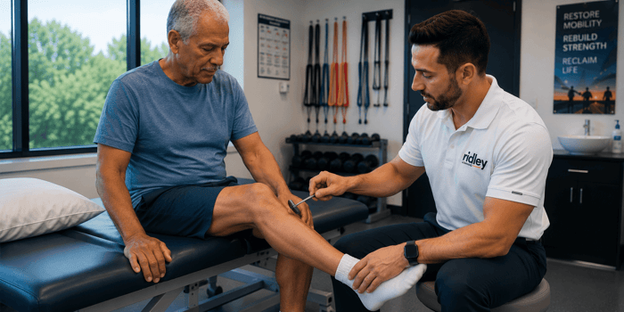
The patient your surgeon is referring out for "PT trial" is the same patient who, in the 2024 literature, has roughly a one-in-two chance of being statistically indistinguishable from a surgical patient six years from now. That's only true if the trial is real. Six visits and a sheet of generic lumbar exercises is not the conservative arm in the trials. Supervised, twice-weekly, multimodal programming for 6 to 12 weeks is.
The non-negotiables: flexion-based exercise (double knee-to-chest, posterior pelvic tilts, seated lumbar flexion progressed to standing), trunk stabilization layered onto that base, and aerobic conditioning in a flexed posture (recumbent or stationary cycling, aquatic work, inclined treadmill) because level walking is the activity that provokes symptoms. Manual therapy as adjunct, not centerpiece. Lumbopelvic mobilization at moderate dose has the strongest dose-response signal.
For Occupational Therapists
What stenosis actually does is interrupt life at the kitchen counter and the laundry basket. The patient whose imaging is severe but whose function is preserved has built compensations you can name from the doorway: shopping cart use, seated tasks, head-of-bed elevation. The patient with mild imaging and severe disability often has not built those compensations.
Energy conservation and activity pacing are not "soft" interventions in this population. They are the primary mechanism by which a stenosis patient stays out of the emergency department and away from the surgical consult. Environment assessment, with attention to standing surface heights and bathroom and kitchen ergonomics, may do more for daily function than the most rigorous strengthening program.
For Athletic Trainers
The tactical and masters populations are where stenosis is now showing up. Load carriage and sustained extension are the loading patterns that break down. The military rehabilitation literature on graded progression from forward-flexed aerobic work back to loaded ambulation translates almost directly to the older athlete returning to ruck marches, ladder work, or even prolonged standing on a sideline.
Cycling preserves cardiovascular base while symptoms persist. Aquatic conditioning reduces axial spinal load by roughly half compared to land walking. Both are appropriate bridges back to land-based and weight-bearing work.
For Massage Therapists
You are often the only manual provider a stenosis patient sees long-term. The most valuable thing you deliver is not the soft tissue work. It is the five minutes after, where you describe what positions open the canal and what positions close it. A client who leaves the table understanding the shopping cart sign has a tool that compounds across years of daily life. Most of them have never had that conversation.
Where the Evidence Is Still Developing
The convergence data, while consistent, mostly captures populations who tolerated their assigned arm. Crossover rates in trials like SPORT were substantial. The patient who fails a real conservative trial and then proceeds to surgery is a different population than either pure arm, and we have less data on them than we should.
Paraspinal multifidus fat as a prognostic variable is replicable but not yet a standard radiology read. Whether prehabilitation specifically targeting multifidus quality changes that prognostic signal is an open question. The Punnoose meta-analysis on orthopedic prehabilitation suggests it might.
And the question of which specific patients should be referred for early surgery (rather than after a conservative trial) is still defined more by red flags (progressive motor deficit, cauda equina symptoms, severe walking limitation under 100 meters) than by a positive predictive rule. The N-CLASS criteria help with diagnosis. A "who-needs-surgery" rule of comparable strength does not yet exist.
Product Spotlight:
Lumbar Spinal Stenosis: Evidence-Based Update

$37.97
This course offers healthcare professionals a comprehensive review of lumbar spinal stenosis, focusing on differential diagnosis, validated assessment criteria, and evidence-based rehabilitation protocols. Participants will learn to differentiate stenosis types, apply clinical reasoning to complex cases, and select appropriate interventions,… read more
Three Things You Can Do This Week
The point of the 2024 evidence isn't a new framework. It's a small number of changes any clinician can make immediately:
- Stop reading the MRI report as a triage tool. Use it to confirm or rule out red-flag pathology and to characterize the location pattern (central, lateral recess, foraminal). The severity grade does not tell you who will respond. Pair every imaging review with N-CLASS criteria and a real physical exam.
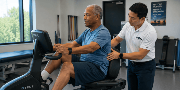
- Make stationary cycling the first aerobic prescription. The patient who cannot walk one block often tolerates 30 minutes of recumbent or stationary cycling without symptom provocation. Use it to break the deconditioning cycle in the first two weeks before progressing to inclined treadmill or aquatic work.
- Define what "adequate conservative trial" means before you start. Six visits and a generic exercise sheet is not the comparator arm in the Nordsten-DS or convergence trials. Supervised, twice-weekly, multimodal for 6 to 12 weeks is. If your scheduling cannot support that, build a hybrid model with monitored home programming and decision benchmarks at weeks 4 and 8.
In a large cohort analysis by Anderson and colleagues across more than 11,000 low back pain episodes, a single visit of spinal manipulation was associated with a 55 percent lower risk of care escalation. Two to twelve visits: 42 percent lower. Thirteen or more visits: no protective effect. Dose matters in a direction most of us don't intuitively expect.
The shared decision-making conversation gets easier when the underlying data is honest. Patients who hear "your imaging is severe, but the severity doesn't predict whether you'll respond to rehab. Here's what we're going to try" engage differently than patients who hear "your imaging is severe, you'll probably need surgery eventually." Both can be true. Only one is supported by the convergence and the imaging-prediction literature.
This is also where Ridley Learning's full evidence stack for this condition lives. The course pulls the Nordsten-DS, Shahidi, Wesselink, and AAN 2025 epidural data into one operating model, with the discipline-specific protocols sitting underneath. The infographic series in the related myths breakdown covers the same evidence in a different format.
The Bottom Line
Imaging severity tells you what the anatomy looks like. It does not tell you who will respond to lumbar spinal stenosis rehabilitation. An adequate supervised multimodal trial is the appropriate first step for almost every symptomatic stenosis patient, and the long-term outcomes meet the surgical outcomes at the finish line. The patient in your clinic next week deserves that conversation before the surgical referral, not after it.
FAQs
Does imaging severity ever justify earlier surgical referral in lumbar spinal stenosis rehabilitation?
Imaging severity alone does not. Progressive neurological deficit, cauda equina symptoms, walking distance under 100 meters that is not improving, or failure of a 6 to 12 week supervised conservative trial are the actual triggers. A severe MRI in a patient who is functioning and motivated is still an indication for an adequate rehabilitation trial first.
How do I structure the conservative trial so I can defend its adequacy to a surgeon or insurer?
Supervised, twice weekly, 6 to 12 weeks, multimodal. Manual therapy, flexion-based exercise, trunk stabilization, aerobic conditioning in flexed posture, and structured patient education. Document outcome measures (Oswestry Disability Index, walking distance, leg pain numeric scale) at baseline, weeks 4 and 8, and discharge. This matches the comparator arm in the convergence and Nordsten-DS trials.
What if my stenosis patient also has obesity, diabetes, and knee osteoarthritis?
Aquatic therapy is the entry point. Pool walking in chest-deep water reduces axial spinal load by approximately 50 percent and unloads the knee simultaneously. Land-based progression follows symptom and capacity tolerance. The comorbidity burden does not preclude meaningful response; it changes the modality you start with. See flexion-based exercise programming for details.
If decompression alone matches decompression plus fusion, why is fusion still being done for spondylolisthesis?
The Nordsten-DS 5-year publication in BMJ (2024) and multiple meta-analyses find no clinical outcome difference. Fusion remains appropriate for high-grade spondylolisthesis or gross instability. For typical degenerative grade 1 spondylolisthesis with stenosis, decompression alone is now the better-supported approach. Practice patterns lag the evidence.
Does the "imaging-doesn't-predict finding" apply to elderly or frail patients too?
Yes. Frailty and high comorbidity burden predict worse baseline function and slower recovery trajectories, but they do not preclude meaningful response to rehabilitation. The trajectory is smaller and slower; the direction of change is the same. This subgroup is the one most often denied an adequate trial, despite having the most to lose from surgical complications.